
What is Basal Cell Skin Cancer?
Basalioma is one of the most common skin tumors (60-80% of all malignant skin tumors). Basalioma is most commonly found over the age of 50 years. Typical localization is the face (forehead, nose, inner corner of the eye, nasolabial fold), neck.
Pathogenesis During Basal Cell Skin Cancer
According to some authors, basal cell carcinoma refers to localized tumors, as it is not a truly malignant tumor. Basalioma has signs characteristic of a malignant process:
- rapid tumor growth with infiltration;
- destruction of underlying tissues;
- recurrence ability (even after properly performed treatment). However, basalioma is a local malignant tumor; it does not metastasize.
Predisposing factors:
- increased insolation;
- ionizing radiation;
- contact with various harmful substances (arsenic, hydrocarbon);
- constant trauma of the skin area.
Pathological anatomy. Bazalioma develops from the cutaneous epidermis. This tumor was built from small cells of oval, round or spindle shape, with a narrow rim of basophilic cytoplasm. These cells resemble in their structure the cells of the basal layer of the skin. Such cells are nests or cords. Basalioma grows slowly, gradually spreading from the depths of the epidermis to the surface of the skin.
Symptoms of Basal Cell Skin Cancer
Clinical forms of basal cell carcinoma
- Nodular ulcerative form (ulcus rodens).
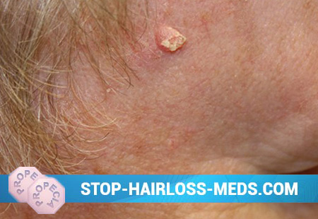
The most frequent localization of this form of basal cell carcinoma is the eyelids, the inner corner of the eye, and nososchechnye folds. On the surface of unchanged skin there is a nodule, dense in texture and protruding above the skin surface. The color of this nodule can be different – from pink to red. The skin above the nodule may be dull or shiny, thinner. Over a long period of time, this nodule begins to ulcerate, grow in size, the bottom of the ulcer is covered with greasy bloom. Gradually, such a nodule acquires irregular shapes, telangiectasias appear on its surface. In the center of the nodule a crust forms over the ulceration. Along the edges of such a knot, a dense roller with rounded pearl-colored edges is formed.
The tumor significantly destroys the surrounding tissue, but never metastasizes.
- Proprietary form.
This form occurs more frequently in areas of the skin that are constantly traumatized. In its clinical picture, this form is similar to the nodular-ulcerative form, but differs from it in more rapid development and more pronounced destructive growth. Prodazy basalioma is rare.
- Warty (exophytic, papillary) form.
This type of tumor is characterized by the fact that its growth is not infiltrating, as in the vast majority of cases, but in the form of “cauliflower”, i.e. the tumor spreads over the skin surface in the form of hemispherical nodes that are dense on palpation and protrude above the skin surface.
- Nodular (large-silk) form.
The difference of this form from ulcus rodens is that nodular basalioma does not grow in the depth of the skin, but upwards. She has the appearance of a hemispherical solitary formation protruding above the surface of the skin; telangiectasia shines through her tissues.
- Pigment form.
This form of basal cell carcinoma must be differentiated from malignant melanoma. All signs of basal cell carcinoma in this nodule are preserved, it has a characteristic appearance and a “pearl” bezel, but in the center or along the edges of the basal cell carcinoma there is brown or black pigmentation.
Cicatricial atrophic form. The onset of tumor development is similar to ulcus rodens – the nodule gradually increases in size, begins to ulcerate, and a flat ulcer with a characteristic “pearl” cushion along the periphery is formed. Then spontaneous cicatrization of this erosion begins, that is, in the center this erosion is scarring, and tumor growth continues along the periphery.
- Sclerodermiform form.
Dakzhe tends to process scarring. Initially, the skin has a small dense pale nodule, then it gradually increases in size – a dense flat plaque with translucent telangiectasias is formed. Subsequently, this plaque may ulcerate.
- Pedzhetoidnaya (surface) form, or pedzhetoidnaya epithelioma, flat surface basal cell carcinoma.
This type of tumor is localized more often in closed areas of the body. Pegetoid epithelioma is a multiple tumor that does not infiltrate the skin and does not rise above its surface. This tumor is of various shades of red (from pale pink to red), flat with characteristic raised pearl-colored edges up to 4 cm. The duration of development of such a tumor over time is large, several decades, is benign. Separate separately syndrome Gorlin – Holtz, which includes the pedzhetoidnuyu form of basal cell carcinoma, multiple cysts of the mandible, anomalies of the ribs and other malformations.
- Shpigler’s tumor (cylinder, “turban” tumor).
This tumor is located on the scalp. Shpigler’s tumor consists of a set of dense nodes of a hemispherical shape of purple-pink color with a broad base, the diameter of the nodes is 1-10 cm. The surface of the tumor is covered with telangiectasias. The development of this tumor for a long time, the process is relatively benign.
Diagnosis of Basal Cell Skin Cancer
The main diagnostic method is cytological (cellular) research. The information content of this technique is ~ 88-98%. Samples – prints and scrapings from the surface of the tumor are examined.
Treatment of Basal Cell Skin Cancer
- surgical treatment (under local anesthesia);
- radiotherapy (close focus, total dose is 30-60 gray);
- cryodestruction (temperature – 180-190 ° С, 1-2 applications are performed);
- local chemotherapy (applications with metatrexate, fluorouracil, colchamin);
- laser coagulation of the tumor. Squamous cell carcinoma There are many synonyms for squamous cell carcinoma – spinocellular carcinoma, spinalioma, epithelioma planocellulare.
Forecast. Basal cell carcinoma is not prone to metastasis. However, it is characterized by frequent recurrence — even after adequate treatment — in 25–80% of cases. Therefore, running forms are considered to be larger than 2 cm in diameter. The prognosis is favorable.